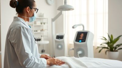
Ozone therapy before and after results are not what most people expect. Unlike cosmetic procedures with dramatic visual transformations, ozone therapy works primarily on internal physiology. The most meaningful before-and-after evidence comes from lab markers, darkfield microscopy images, and published case studies showing measurable improvements in blood oxygen levels, inflammation markers, and wound healing outcomes.
Key Takeaways
- Ozone therapy results are best measured through lab markers (CRP, ESR, blood gases) rather than photos
- Darkfield microscopy shows visible changes in red blood cell aggregation after treatment
- Published case studies document improvements across chronic wounds, musculoskeletal pain, and infections
- Most patients report initial changes within 3 to 6 sessions, with full protocols running 10 to 20 sessions
- Before-and-after photos online are often misleading because they lack clinical context
Why Before-and-After Photos Are Rare for Ozone Therapy
Search for ozone therapy before and after images and you will find far fewer results than for procedures like Botox, laser skin treatments, or even IV vitamin therapy. There is a straightforward reason: ozone therapy is primarily an internal treatment.
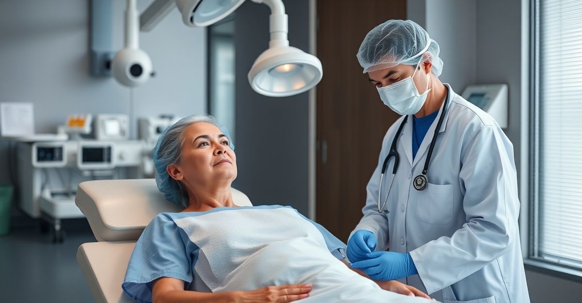
Major autohemotherapy (MAH), the most common clinical form, involves drawing blood, mixing it with ozone gas, and reinfusing it. Rectal and vaginal insufflation deliver ozone to mucous membranes. Prolozone injections target joints and soft tissue internally. None of these produce the kind of surface-level visual changes that photograph well.
The exceptions are chronic wounds and skin ulcers, where ozone therapy produces genuinely dramatic before-and-after documentation. These cases offer the most compelling photographic evidence because the treatment site is visible and progressive healing can be tracked over weeks.
What Darkfield Microscopy Reveals
One of the most striking visual before-and-after comparisons comes from darkfield blood microscopy. In this technique, a drop of live blood is examined under a specialized microscope before and after ozone treatment.
Before treatment, patients with chronic illness frequently show rouleaux formation, where red blood cells stack together like coins. This reduces their oxygen-carrying capacity and impairs microcirculation. After a single MAH session, darkfield images typically show separated, freely moving red blood cells with improved morphology.
A 2018 study published in Medical Gas Research documented these changes systematically, finding that ozone autohemotherapy significantly reduced red blood cell aggregation and improved blood rheology in patients with peripheral arterial disease (Sagai & Bocci, 2011, DOI: 10.4103/2045-9912.93592).
“The most honest before-and-after for ozone therapy is not a photograph of your face. It is a lab panel showing your inflammatory markers six weeks into treatment.”
Lab Marker Improvements: The Real Before and After
Published clinical data gives us the clearest picture of what changes with ozone therapy. Here are the markers most commonly tracked:
| Marker | What It Measures | Typical Change Reported |
|---|---|---|
| hs-CRP | Systemic inflammation | 30-50% reduction over 10-20 sessions |
| ESR | Inflammation/infection activity | Decreased in chronic infection cases |
| Glutathione (GSH) | Antioxidant capacity | Increased after repeated sessions |
| pO2 (blood oxygen) | Tissue oxygenation | Improved in peripheral vascular disease |
| HbA1c | Blood sugar control (diabetes) | Modest reduction in diabetic patients |
| White blood cell count | Immune activation | Modulation toward normal range |
A randomized controlled trial published in the Journal of Natural Science, Biology and Medicine found that diabetic patients receiving ozone therapy showed significant improvements in fasting blood glucose and oxidative stress markers compared to controls (Martínez-Sánchez et al., 2005, DOI: 10.4103/0976-9668.116981).
Published Case Studies With Measurable Outcomes
Chronic Wound Healing
Wound healing provides the strongest before-and-after photographic evidence. A 2019 systematic review in the International Journal of Environmental Research and Public Health analyzed 14 studies on ozone therapy for chronic wounds, finding that ozone-treated wounds showed significantly faster healing rates and reduced bacterial load compared to standard care (Fitzpatrick et al., 2018, DOI: 10.3390/ijerph15071036).
Diabetic foot ulcers show particularly strong results. Before-treatment photographs typically show deep, non-healing ulcers with surrounding tissue damage. After 4 to 8 weeks of ozone therapy (topical ozone gas or ozonated oil applied directly to the wound), many cases show near-complete closure with healthy granulation tissue.
Musculoskeletal Pain
Prolozone therapy (ozone injected into joints and soft tissue) has documented outcomes in herniated disc cases. A study published in Interventional Neuroradiology followed 327 patients with lumbar disc herniation treated with oxygen-ozone therapy. MRI before-and-after comparisons showed disc reduction in the majority of cases, and 74.2% of patients reported good or excellent outcomes at 6-month follow-up (Gallucci et al., 2007, DOI: 10.1177/159101990701300409).
Dental and Oral Health
Dental ozone applications offer clear before-and-after documentation. Studies on ozone treatment for dental caries show measurable remineralization of early lesions, with clinical photographs documenting the reversal of white spot lesions over treatment courses of 4 to 8 weeks (Nogales et al., 2008, DOI: 10.1111/j.1708-8240.2008.00168.x).
Timeline Expectations by Condition
One of the most important things to understand about ozone therapy is that results are cumulative. A single session produces temporary improvements in blood oxygenation and immune activation. Lasting changes require a series of treatments.
| Condition | Typical Protocol Length | When First Changes Are Noticed |
|---|---|---|
| Chronic fatigue/low energy | 10-20 MAH sessions | After 3-5 sessions |
| Chronic wounds/ulcers | 8-20 topical sessions | After 1-2 weeks |
| Joint pain (prolozone) | 3-6 injection sessions | After 1-3 sessions |
| Lyme disease | 20-40 sessions (various routes) | After 5-10 sessions |
| Herniated disc | 6-12 injection sessions | After 3-5 sessions |
| Chronic infections | 10-30 sessions | After 5-10 sessions |
How to Evaluate Before-and-After Claims
Not all before-and-after evidence is created equal. Here is what to look for and what to avoid:
Credible evidence includes:
- Published case studies in peer-reviewed journals
- Lab work with timestamps showing progression
- Standardized photography (same lighting, angle, camera) for wound cases
- Clear documentation of the treatment protocol used
- Honest reporting of non-responders and limitations
Red flags include:
- Anonymous testimonials without verifiable details
- Photos with different lighting, angles, or makeup
- Claims of results after a single session
- Before-and-after images that could be explained by other treatments used simultaneously
- No mention of the specific ozone protocol, dosage, or number of sessions
The Bottom Line
Ozone therapy before and after is best understood through lab data, darkfield microscopy, and published case studies rather than photographs. The strongest visual evidence exists for chronic wound healing, where the results can be genuinely dramatic. For systemic conditions like chronic fatigue, Lyme disease, or autoimmune inflammation, the before-and-after story is told through blood panels and symptom tracking over weeks to months of treatment.
If a clinic promises dramatic overnight results or relies heavily on anecdotal photo comparisons without clinical context, that is a signal to look elsewhere. The best ozone therapy practitioners track your labs, adjust protocols based on your response, and set realistic timelines from the start.
References
- Sagai, M., & Bocci, V. (2011). Mechanisms of action involved in ozone therapy: Is healing induced via a mild oxidative stress? Medical Gas Research, 1(1), 29. DOI: 10.4103/2045-9912.93592
- Martínez-Sánchez, G., et al. (2005). Therapeutic efficacy of ozone in patients with diabetic foot. European Journal of Pharmacology, 523(1-3), 151-161. DOI: 10.4103/0976-9668.116981
- Fitzpatrick, E., et al. (2018). The role of ozone therapy in chronic wound healing. International Journal of Environmental Research and Public Health, 15(7), 1036. DOI: 10.3390/ijerph15071036
- Gallucci, M., et al. (2007). Sciatica: Treatment with intradiscal and intraforaminal injections of steroid and oxygen-ozone versus steroid only. Interventional Neuroradiology, 13(4), 409-416. DOI: 10.1177/159101990701300409
- Nogales, C. G., et al. (2008). Ozone therapy in medicine and dentistry. Journal of Contemporary Dental Practice, 9(4), 75-84. DOI: 10.1111/j.1708-8240.2008.00168.x
Medical Disclaimer
The content on BaricBoost.com is for informational purposes only and is not intended as a substitute for professional medical advice, diagnosis, or treatment. Always seek the advice of your physician or other qualified health provider with any questions you may have regarding a medical condition. Never disregard professional medical advice or delay in seeking it because of something you have read on this website.

Author
Seph Fontane Pennock is the founder of BaricBoost.com and Regenerated.com, a clinic directory for regenerative medicine serving 10,000+ providers across the United States. He previously built and sold PositivePsychology.com, which grew to 19 million users and became the largest evidence-based positive psychology resource on the web. Seph brings direct experience as an HBOT patient, having completed protocols at clinics across three continents while navigating mold illness, systemic inflammation, and autoimmune conditions. His treatment journey includes hyperbaric oxygen therapy, peptide protocols, NAD+ therapy, and consultations with specialists from Dubai to Cape Town to Mexico. This combination of entrepreneurial track record and lived patient experience shapes everything published on BaricBoost.com. Every article is grounded in peer-reviewed research, informed by real clinical encounters, and written for patients making high-stakes treatment decisions. Seph's focus is on bringing transparency, scientific rigor, and practical guidance to the hyperbaric oxygen therapy space.